Methods for purifying and analysing proteins via western blotting

For many years, the western blotting technique has been a primary method in molecular biology and techniques like proteomics (the large-scale study of proteomes: a set of proteins produced in an organism, system, or biological context). Given that western blot is a multistep process, often requires specialist interpretation and potential errors and variations occurring at any step can compromise the reliability and reproducibility of its results.
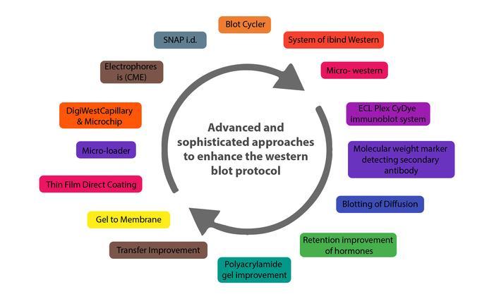
Given this situation, in the last two decades, many advanced and sophisticated approaches have been developed to improve the western blot protocol. These improvements are more automated and sensitive, with high capabilities in reducing potential issues with the traditional western blot method. The enhanced sensitivity and innovative equipment advancements in western blotting can expand the field of the clinical applications of this foundational method.
In this article, Helvetica Health Care tells you all about the western blot technique as a method of purifying and analysing proteins and the new developments in western blotting methodology.
What is the Western blot technique ?
Traditionally, the western blotting method involves seven steps, as below.
- Sample preparation from cells or tissue lysates
- Separation of proteins by gel electrophoresis
- Protein transfer in a nitrocellulose or polyvinylidene fluoride (PVDF) membrane,
- Blocking of non-specific proteins on membrane
- Primary Antibody incubation
- Secondary Antibody incubation
- Protein detection & analysis
What are the new developments in the western blot domain and what are their characteristics?
Below are some innovations that have enhanced the western blot protocol and addressed the potential problems in its application in protein purification and analysis.
Capillary and microchip electrophoresis (MCE)
MCE provides higher sensitivity and better resolution than conventional techniques, enabling the detection of several target proteins from a single cell lysate sample. Besides allowing parallel multiplexed tests of a set of proteins using a small sample amount, the method also eliminates blocking stages and has quicker analysis times (8 min for electrophoretic resolution). Although it’s still developing, MCE could be significantly improved with additional work.
Automated microfluidic protein immunoblotting
Automated protein immunoblotting is a controllable and programmable technology that combines PAGE (polyacrylamide gel electrophoresis) and blotting in a single device. It saves time, avoids several test stages, and reduces the amount of equipment and reagents needed. This technology is cost-effective and reduces the use of reagents since it can identify free prostate-specific antigens in a sample of human seminal fluid in less than 5 minutes. The method is continuously being developed to increase sensitivity and enable protein quantification.
Single cell-resolution western blotting
This highly sensitive method has also been used to analyse protein variations in stem cell signaling and differentiation. It can identify specific cell-to-cell changes in protein expression between cells. The first Single-Cell Western platform in the world, MiloTM (ProteinSimple), can measure protein expression (up to 4 proteins per cell simultaneously) in 1000 single cells in around 4 hours.
DigiWest
DigiWest, a simplified version of the standard procedure, optimises the throughput of western blotting by requiring less lysate, target detection, and antibody. This method, however, calls for the biotinylation of the target proteins, necessitates specialised reagents and equipment, incurs initial startup costs, issues about the translational of the digital DigiWest result to western blots mimics, and specifics about some procedures, like the complex and difficult elution of proteins from PVDF membranes.
Simple Western
Developed by ProteinSimple (San Jose, California), Simple Western is based on CE-SDS (Capillary electrophoresis sodium dodecyl sulphate), wherein the separated proteins are connected to the capillary wall by a patented photo- (ultraviolet) induced chemical crosslink. The method can be performed using 24-90 distinct antibodies simultaneously, is reasonably quick, and uses modest sample amounts. It can also carry out standard curve quantification similar to that done using high-pressure liquid chromatography (HPLC) and enhance reproducibility. However, it needs specialised reagents.
Micro-loader
The western blot assay and PAGE have increased sensitivity thanks to the Micro-loader technology. It uses a sample micro-loader device with a funnel-like construction to load samples. Because it only needs a minimal number of loading samples for signal detection, this technique is highly effective for measuring protein expression and phosphorylation in samples that are few, hard to find, and limited in quantity.
Thin-film direct coating with suction-western blotting (TDCS)
High antibody consumption and lengthy operating times are major limitations of western blotting. TDCS, a capillary-tube-based technique, aims to cut down on antibody and time consumption for western blotting. This quick and sensitive detection method allows for the quantitative study of multiple antigen-antibody interactions and can do the same for multiple protein interactions.
Blotting of diffusion
SDS-PAGE single-prefabricated gels on plastic assistance are a quick and easy approach to making numerous blots using diffusion blotting. Diffusion blotting allows the evaluation of several proteins on blots of the same gel comparable to one another. Protein transmission rates by diffusion blotting are approximately 10% in 3 min, 20% in 20 min, and 40% to 60% in 3 h. The technique may increase the amount of proteins efficiently delivered to the membrane surface, particularly when compared to electroblotting. And so, diffusion blotting has been the preferred method when quantitative protein exchange is not required.
Micro western
Soon, micro western will be a proper western blot technique for confirming the diagnosis of purified HIV p24 and gp120 proteins in blood plasma. The Micro-Western may be vital for detecting infectious diseases and cancer, despite not being widely available.
System of iBind western
Thermo Fisher Scientific’s iBind Western procedure employs a low-cost semi-automated western blotting structure that leverages sequential flow technology to distribute chemicals and antibodies to various compartments. Using the iBind Western Systems and highly specialised primary and secondary antibodies will result in cleaner western blots.
Blot cycler
With the BlotCycler (Precision Biosystems, USA), every step of the membrane processing is automated, and up to 12 membranes can be processed at once. The BlotCyclerTM is an Automated Western Blot Development equipment that completes all blot cleaning, trapping, and incubation steps with improved repeatability and sensitivity. It also is simple to programme all the actions for particular operations.
System of SNAP i.d.
In a low-cost system of SNAP i.d. 2.0, solvents are uniformly distributed via the tissue using a technique based on a vacuum and a flow distribution system (Merck, USA). Its main advantage is that washing, antibody incubation, and blocking all take about 30 minutes. The SNAP i.d.® 2.0 gadget actively forces reagents through the membrane, unlike conventional Western blotting, which depends on diffusion to transfer reagents.
Washes, antibody recollection, and antibody-antigen binding have been improved. The SNAP i.d.® 2.0 delivers new, cutting-edge western blotting characteristics with an immunodetection capability in 30 minutes without using additional reagents (e.g., antigen, antibody, or detection reagents).
Retention improvement of hormones of the peptide on the membrane of blotting
Western blotting is significantly improved by treating the PVDF membrane used in this method with glutaraldehyde (0.2%) for fifteen minutes in a saline solution containing tween-80. In western blotting, the addition of glutaraldehyde to the membrane prevented or reduced the quantity of insulin loss.
Apart from these, two more techniques that have improved the conventional method are
- Analysis of western blot utilising molecular weight marker detecting secondary antibody
- Total and target protein co-detection by immunoblotting of fluorescent ECL and labelling of Cydye
Many innovations have also been observed in western blotting techniques to transfer proteins from gel to membrane, such as Vacuum blotting, Centrifuge blotting, Multiple tissue blotting, etc. Improvements in protein transfer methods from gels to membranes include the Trans-Blot Turbo, the iBlot® dry blotting system, and Pierce™ Power Blot Cassette. When these new techniques are used, the transfer time is reduced from an hour or overnight to at most 10 minutes.
HHC offers the ZeptoMetrix WESTERN BLOT for in vitro detection of antibodies to SIV in serum or plasma and is available in 10 or 30-strip kit formats. We also provide a bench top, a western blot processor, and the AUTOBLOT 3000. It controls incubation times, dispensing volumes and washing programs. Ten user-defined protocols can be programmed easily. Contact us now to know how we can help you get accurate and high-quality output with our products when performing the western blotting procedure.